Anatomy Pictures Of Lower Back And Hip - Sacroiliac Joint Syndrome - Yanni : A basic understanding of the anatomy of your lower back can help you identify and differentiate a problem that commonly.
Anatomy Pictures Of Lower Back And Hip - Sacroiliac Joint Syndrome - Yanni : A basic understanding of the anatomy of your lower back can help you identify and differentiate a problem that commonly.. The lumbar region of the spine, more commonly known as the lower back, is situated between the thoracic, or chest, region of the spine, and the sacrum. —the hip bones are largely covered with muscles, so that only at a few points do they approach the surface.she is a former american college of sports medicine certified personal trainer and currently works as a level 1 crossfit. The sacroiliac (si) joints connect the sacrum at the base of the spine with the hip bone. These sections are cervical (neck), thoracic (upper and middle back), lumbar (lower back), and sacrum (tailbone). Anatomy pictures of lower back and hip :
Understanding lower back anatomy 1 the your lower back (lumbar spine) is the anatomic region between your lowest rib and the upper part of the 13.04.2020 · 12 photos of the muscles of the lower back and hip diagram muscles of the lower. These sections are cervical (neck), thoracic (upper and middle back. Understanding lower back anatomy is key to understanding the root of lower back and hip pain. It's also the largest joint in the body. Pain that originates elsewhere may radiate to.
The Skeletal System The Hip And Lower Spine Stock Photo ... from media.istockphoto.com Muscle anatomy neck 12 photos of the muscle anatomy neck dog neck muscle anatomy, front neck muscle anatomy, muscle anatomy neck, muscle anatomy of neck and shoulder, neck muscle anatomy chart, human muscles, dog neck muscle anatomy, front neck muscle anatomy, muscle anatomy neck, muscle anatomy of neck and. Understanding the anatomy of your lower spine can help you communicate more effectively with the medical professionals who treat your lower back pain. Which bones fuse to make the hip and wh… lower limb anatomy. The human spine is composed of 4 sections of vertebrae. It's also the largest joint in the body. It is the muscles of the hip that allow the movements of the hip: Hip pain may result from inflammation, degeneration, or injury to structures and tissues within the hip joint. Check out anatomy picture on ebay.
Those with tight hip flexors.
Hip pain may result from inflammation, degeneration, or injury to structures and tissues within the hip joint. Those with tight hip flexors. Muscle anatomy neck 12 photos of the muscle anatomy neck dog neck muscle anatomy, front neck muscle anatomy, muscle anatomy neck, muscle anatomy of neck and shoulder, neck muscle anatomy chart, human muscles, dog neck muscle anatomy, front neck muscle anatomy, muscle anatomy neck, muscle anatomy of neck and. Picture tests in practical anatomy. Understanding lower back anatomy is key to understanding the root of lower back and hip pain. The muscles of the lower back help stabilize, rotate, flex, and extend the spinal column, which is a bony tower of 24 vertebrae that gives the body structure and houses the spinal cord. The anatomy of the hip and back is comprised of numerous parts that can be injured or wear out, and many problems that occur in this area can display the exact same symptoms or pathology. These muscles provide posture and stability to the body by holding the vertebral column erect and adjusting the position of the body to maintain balance. When most people mention their back, what they are actually referring to is their spine. Understanding lower back anatomy is key to understanding the root of lower back and hip pain. The hip muscles are part of the core. The sacroiliac (si) joints connect the sacrum at the base of the spine with the hip bone. Get anatomy picture with fast and free shipping for many items on ebay.
When most people mention their back, what they are actually referring to is their spine. The muscles of the lower back, including the erector spinae and quadratus lumborum muscles, contract to extend and laterally bend the vertebral column. Understanding lower back anatomy is key to understanding the root of lower back and hip pain. Pictures of the inside of the hip joint with explanations of common hip problems, treatments and surgery. Picture tests in practical anatomy.
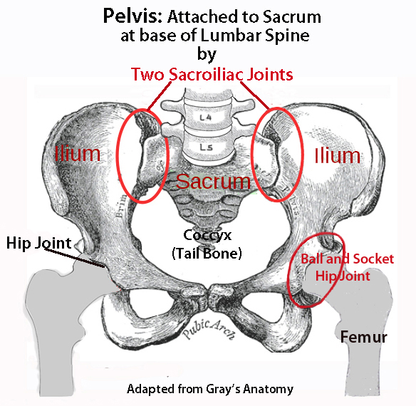
Hip Joint Anatomy | Bone and Spine from boneandspine.com The human spine is composed of 4 sections of vertebrae. This article looks at the anatomy of the. It is the muscles of the hip that allow the movements of the hip: —the hip bones are largely covered with muscles, so that only at a few points do they approach the surface.she is a former american college of sports medicine certified personal trainer and currently works as a level 1 crossfit. The vertebral column of the lower back includes the five lumbar vertebrae, the sacrum, and the coccyx. Those with tight hip flexors. Anatomy pictures of lower back and hip : The hip region is located lateral and anterior to the gluteal region, inferior to the iliac crest, and overlying the greater trochanter of the femur, or thigh bone.
Those with tight hip flexors.
Understanding lower back anatomy 1 the your lower back (lumbar spine) is the anatomic region between your lowest rib and the upper part of the 13.04.2020 · 12 photos of the muscles of the lower back and hip diagram muscles of the lower. It's also the largest joint in the body. When most people mention their back, what they are actually referring to is their spine. The anatomy of the hip and back is comprised of numerous parts that can be injured or wear out, and many problems that occur in this area can display the exact same symptoms or pathology. Possible causes of lower back and hip pain include sprains, strains, and a herniated disk. Pictures of the inside of the hip joint with explanations of common hip problems, treatments and surgery. This article looks at the anatomy of the. When something injures or puts pressure on the sciatic nerve, it can cause pain in the lower back that spreads to the hip, buttocks, and leg. These sections are cervical (neck), thoracic (upper and middle back. It is the muscles of the hip that allow the movements of the hip: Which bones fuse to make the hip and wh… lower limb anatomy. The hip region is located lateral and anterior to the gluteal region, inferior to the iliac crest, and overlying the greater trochanter of the femur, or thigh bone. The bones of the pelvis and lower back work together to support the body's weight, anchor the abdominal and hip muscles, and protect the delicate vital organs of the vertebral and abdominopelvic cavities.
The bones of the pelvis and lower back work together to support the body's weight, anchor the abdominal and hip muscles, and protect the delicate vital organs of the vertebral and abdominopelvic cavities. Pain that originates elsewhere may radiate to. When a person experiences lower back and hip pain simultaneously, there may be an underlying injury or medical. Abdominal muscle anatomy pictures of abdominal muscles. The muscles of the lower back help stabilize, rotate, flex, and extend the spinal column, which is a bony tower of 24 vertebrae that gives the body structure and houses the spinal cord.
The Rectus Femoris and Lower Back Pain from corewalking.com Anatomy pictures of lower back and hip : The bones of the pelvis and lower back work together to support the body's weight, anchor the abdominal and hip muscles, and protect the delicate vital organs of the vertebral and abdominopelvic cavities. Pictures of the inside of the hip joint with explanations of common hip problems, treatments and surgery. Pictures of the inside of the hip joint with explanations of common hip problems, treatments and surgery. A basic understanding of the anatomy of your lower back can help you identify and differentiate a problem that commonly. Picture tests in practical anatomy. When a person experiences lower back and hip pain simultaneously, there may be an underlying injury or medical. The vertebral column consists of 33 vertebrae which can be split up into 5 continuous sections.
Muscle anatomy neck 12 photos of the muscle anatomy neck dog neck muscle anatomy, front neck muscle anatomy, muscle anatomy neck, muscle anatomy of neck and shoulder, neck muscle anatomy chart, human muscles, dog neck muscle anatomy, front neck muscle anatomy, muscle anatomy neck, muscle anatomy of neck and.
These sections are cervical (neck), thoracic (upper and middle back. But did you check ebay? Anatomy pictures of lower back and hip : The hip joint is the uppermost part of the leg where the head of the thigh bone (femur) fits into the socket of the pelvis. Pain that originates elsewhere may radiate to. Understanding lower back anatomy is key to understanding the root of lower back and hip pain. The muscles of the lower back, including the erector spinae and quadratus lumborum muscles, contract to extend and laterally bend the vertebral column. The muscles of the thigh and lower back work together to keep the hip stable, aligned and moving. This article looks at the anatomy of the. It's also the largest joint in the body. Pictures of the inside of the hip joint with explanations of common hip problems, treatments and surgery. The human spine is composed of 4 sections of vertebrae. Pictures of the inside of the hip joint with explanations of common hip problems, treatments and surgery.


0 Comments